Retinal imaging & OCT
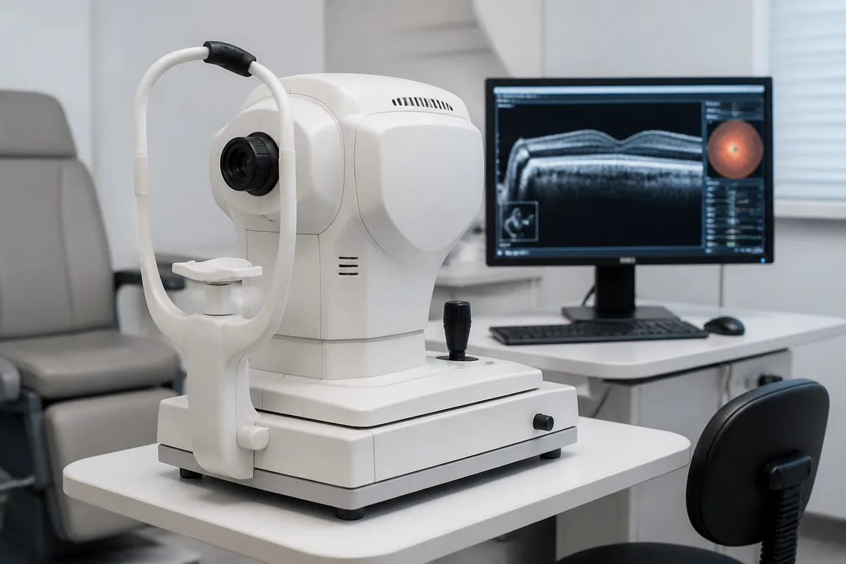
Retinal imaging & OCT
Optical coherence tomography produces a cross-section of the retina at microscopic resolution, showing layers that no view through a lens can reveal. Combined with widefield retinal photography, it is how we catch disease before it affects your sight.
What's Involved
- Widefield retinal photography — A broad, documented image of the retina without the wait for dilation.
- OCT cross-sections — Layer-by-layer measurement of the macula and optic nerve.
- Year-on-year comparison — Scans stored and compared so real change is distinguished from normal variation.
When to Come In
- Diabetes or high blood pressure — Conditions that damage the retina silently.
- Glaucoma risk or suspicion — Nerve fibre thinning shows on OCT years before field loss.
- Any unexplained vision change — Blur, distortion, or a spot that a refraction does not explain.
This content is for general information only and isn't medical advice. Results vary from person to person, so talk with your eye doctor about your own eyes. If you have a medical emergency, call 911.